Endoscope Diameter and Foraminal Access in Endoscopic Spine Surgery

Introduction: Why Endoscope Diameter Matters

Endoscope diameter is not a minor technical detail in endoscopic spine surgery. It directly affects whether the natural lumbar foraminal corridor is sufficient—or whether bony removal becomes necessary.

In transforaminal endoscopic approaches, safety is determined by the interaction between foraminal anatomy, the working cannula diameter, and the required surgical trajectory. Understanding that relationship helps optimize nerve safety and preserve facet integrity.


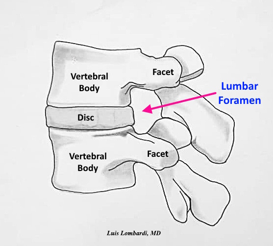
Endoscope Diameter and the Foraminal Working Corridor

The lumbar foramen offers a limited surgical corridor. The usable working space varies with:

  • Disc height
  • Facet orientation
  • Foraminal dimensions
  • Degenerative narrowing
  • Endoscope and cannula diameter

As instrument diameter increases, the corridor required for safe passage increases. When native space is insufficient, corridor expansion—often through facet resection—may be required.

By contrast, smaller-diameter endoscopes can work within tighter corridors while preserving native bony anatomy.

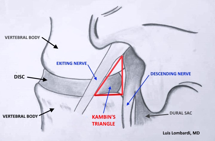
Working Cannula Diameter, Scope Size, and 3D Foraminal Constraints

The transforaminal working corridor is a three-dimensional space, not a flat triangle. With increasing cannula diameter:

  • Clearance within Kambin’s triangle decreases.
  • The risk of proximity to (or compression of) the exiting nerve root increases.
  • The superior articular process becomes a dominant limiting boundary in the posterolateral corridor.
  • The margin of safety narrows.

Even small differences in cannula size can meaningfully change the trajectory required to maintain safe neural distance. Instrument selection should therefore match patient-specific foraminal anatomy, not habit.


Endoscope Diameter, Facet Removal, and Safe Transforaminal Access

Facet removal is considered when:

  • The foraminal corridor cannot safely accommodate the chosen cannula
  • Degenerative hypertrophy narrows posterolateral access
  • The planned trajectory risks neural contact or compression
  • The target pathology cannot be reached without enlarging the corridor

Larger endoscopic systems require facet removal to create adequate clearance. Smaller systems fit even in narrowed corridors without bone removal.

The decision is anatomical—not procedural.


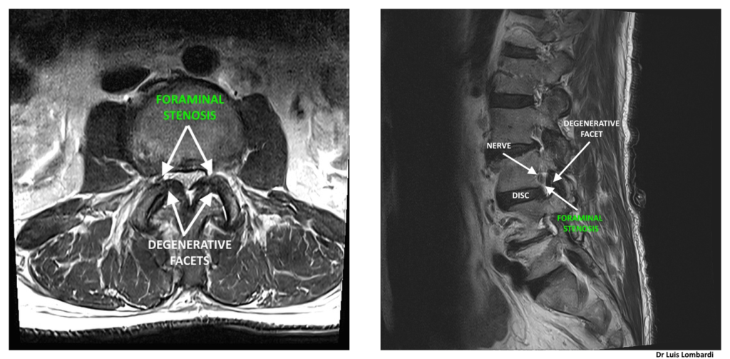
Degenerative Foraminal Stenosis: Choosing Endoscope Diameter and Cannula Size

Degeneration changes both corridor size and feasibility:

  • Disc height loss reduces vertical clearance
  • Facet arthrosis narrows posterolateral access
  • Osteophytes and hypertrophy can constrain trajectory options

In advanced degeneration, selecting a smaller endoscopic system allow safer access while preserving spinal stability and reducing destabilization risk.

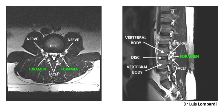
Imaging Planning: Matching Endoscope Diameter to Foraminal Anatomy

Preoperative MRI and CT can help evaluate:

  • Available foraminal width and height
  • Degree and location of facet hypertrophy
  • Nerve root–disc relationship
  • The most realistic working trajectory

Whenever possible, instrument selection should follow objective anatomical assessment rather than preference. Planning based on measurements can reduce unnecessary bone removal and support tissue preservation.

Preservation Strategy: Smaller Endoscope Diameter vs Larger Systems

The objective in lumbar transforaminal endoscopic surgery is not merely to enter the foramen. The goal is to reach the pathology—disc, foramen, or canal—with maximal preservation of normal structures.

Choosing the right endoscope diameter can:

  • Reduce the likelihood of facet removal
  • Minimize destabilization risk
  • Maintain anatomical integrity
  • Support postoperative recovery

Endoscope diameter is therefore a strategic variable in surgical planning, not a footnote.

Conceptual Comparison: Smaller vs Larger Endoscopic Systems (Transforaminal Access)

Table comparing endoscope diameter systems: foraminal clearance, facet work, and degenerative stenosis considerations

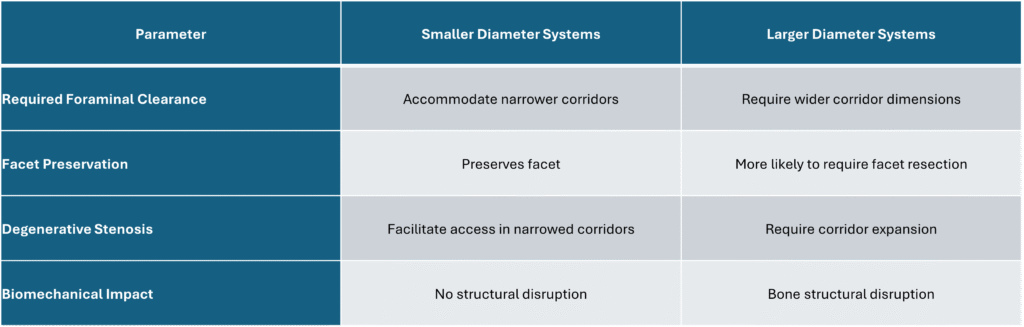
Table 1. Scope Size and Foraminal Access: Operative Considerations


Frequently Asked Questions: Endoscope Diameter and Foraminal Endoscopic Surgery

Does endoscope diameter determine foraminal access?

Larger systems are more likely to require partial facet resection when the natural foraminal corridor is limited.

Can smaller endoscope diameter preserve the foraminal corridor?

Yes. Smaller-diameter systems allow access through narrower corridors while preserving facet anatomy.

How do surgeons decide which endoscope size to use?

Based on imaging, available foraminal dimensions, the planned trajectory, and the degree of degenerative narrowing affecting access.

Internal links

To understand why corridor size varies so much between patients, also see:Lumbar foraminal anatomy

Conclusion: Endoscope Diameter and Foraminal Endoscopic Surgery

In transforaminal endoscopic spine surgery, endoscope diameter and lumbar foraminal anatomy together determine whether facet removal is necessary. Smaller systems enable preservation of native anatomy, while larger systems require corridor expansion through bone resection.

Best outcomes come from individualized anatomical assessment, deliberate trajectory planning, and selecting instrument diameter to match the patient’s foraminal constraints.

Degenerative Foraminal Stenosis and Surgical Planning in Endoscopic Spine Surgery

Introduction

Degenerative foraminal stenosis is an important consideration in endoscopic spine surgery. Progressive anatomical changes within the lumbar foramen can significantly alter the available surgical corridor used during transforaminal procedures.

Successful surgical planning requires a detailed understanding of how degeneration modifies foraminal dimensions, neural relationships, and instrument feasibility.

Why scope size matters: In endoscopic spine surgery, the instruments must fit through the natural opening of the foramen. When the foramen becomes tight from arthritis and disc collapse, smaller endoscopes may allow access with no bone removal, while larger systems require bone removal just to create room for safe insertion.


Pathophysiology of Degenerative Foraminal Stenosis

Degenerative foraminal stenosis develops through a combination of structural changes:

  • Loss of disc height
  • Facet joint hypertrophy
  • Osteophyte formation
  • Ligamentous thickening
  • Segmental instability

As disc height decreases, the vertical dimension of the lumbar foramen is reduced. Simultaneously, facet overgrowth narrows the posterolateral corridor, further compromising the safe working zone.

These progressive changes diminish both neural space and surgical access.


Anatomical Consequences Within the Lumbar Foramen

The lumbar foramen is a dynamic structure rather than a static opening. Degeneration produces:

  • Reduced vertical clearance
  • Decreased horizontal width
  • Altered nerve root orientation
  • Increased neural compression risk

These changes directly impact the geometry of the transforaminal corridor and may distort the classical boundaries described in Kambin’s triangle.

Therefore, degenerative stenosis affects not only symptoms but also surgical strategy.

Lumbar MRI showing a normal foramen with preserved space around the exiting nerve root
Figure 1. Axial (left) and sagittal (right) MRI images demonstrating the lumbar foramen and adjacent anatomical landmarks, including the intervertebral disc, facet joints and exiting nerve root.

These axial and sagittal MRI views show the lumbar foramen and its normal corridor dimensions, which form the anatomical basis for transforaminal endoscopic access planning.

Lumbar MRI showing degenerative foraminal stenosis, where the foramen is narrowed and the exiting nerve root has less surrounding space
Figure 2. Axial (left) and sagittal (right) MRI views illustrating degenerative lumbar foraminal stenosis, with facet hypertrophy and reduction of the foraminal corridor at the involved level.

These views illustrate degenerative foraminal stenosis with narrowing of the foraminal corridor, which directly impacts endoscope diameter selection.


Impact on Endoscopic Transforaminal Access

In patients with degenerative foraminal stenosis, the natural surgical corridor may be substantially narrowed. Narrowing directly affects the geometry of Kambin’s triangle.

This influences:

  • Endoscope diameter selection
  • Cannula positioning
  • Surgical trajectory
  • Need for facet resection

Instrument size can change the plan. Larger-diameter systems may require access-enabling bone removal to create enough room to insert the endoscope safely. Smaller endoscopic systems pass through restricted corridors without bone removal while still treating the pathology.

The choice must be guided by anatomical constraints rather than procedural preference.


Role of Preoperative Imaging

Preoperative MRI and CT imaging are essential in cases of degenerative foraminal stenosis.

Imaging allows evaluation of:

  • Degree of disc collapse
  • Facet hypertrophy severity
  • Foraminal width
  • Osteophyte encroachment
  • Neural compression

Imaging also helps the surgeon estimate whether the corridor can accommodate the planned endoscope size.

Individualized planning is particularly critical in advanced degeneration.


Surgical Strategy: Preservation

The surgical objective is to achieve adequate treatment of the pathology while preserving spinal stability.

Decision-making includes:

  • Selecting an endoscope size that matches the available corridor
  • Using the smallest effective system when anatomy is tight, to reduce the need for bone removal
  • Avoiding unnecessary bone removal that could affect stability

Precision planning minimizes risk and optimizes outcomes.

Conclusion

Degenerative foraminal stenosis significantly alters lumbar foraminal anatomy and directly influences surgical planning in endoscopic spine procedures.

Understanding the anatomical consequences of disc collapse and facet hypertrophy is essential for selecting instrument size and planning trajectory.


Frequently Asked Questions About Degenerative Foraminal Stenosis

What causes degenerative foraminal stenosis?

It results from age-related structural changes such as disc height loss, facet arthritis, and osteophyte formation that progressively narrow the lumbar foramen.

Does degenerative stenosis always require facet removal?

Not always. The need for facet resection depends on the degree of narrowing and the required surgical corridor. Careful imaging evaluation determines the appropriate approach. Smaller endoscopes do not require facet removal, even in degenerative foraminal stenosis.

Can a smaller endoscope reduce the need for bone removal?

Yes. When the foramen is narrowed, a smaller endoscope may fit through the available space with no bone trimming.


More on Lumbar Foraminal Anatomy

Kambin’s Triangle: Anatomical Boundaries and Surgical Implications in Endoscopic Spine Surgery

Introduction

Kambin’s triangle is a key anatomical landmark in endoscopic spine surgery and represents a critical component of the lumbar foramen used during transforaminal access. It is a corridor to access the disc and the spinal canal. Understanding its boundaries, anatomical variability, and surgical limitations are essential for safe and effective endoscopic spine surgery.

Although traditionally described as a triangular safe zone, its dimensions are not fixed. Patient-specific anatomical variations and degenerative changes significantly influence the true working space available during surgery.


Kambin’s Triangle endoscopic surgery boundaries

Kambin’s triangle is defined by three primary anatomical structures:

  • Hypotenuse: Exiting nerve root
  • Base: Superior endplate of the caudal vertebra
  • Height: Descending nerve root

This triangular corridor provides a pathway to the posterior disc and the spinal canal without direct retraction of neural elements when appropriate technique and trajectory are used.

However, the shape may be more trapezoidal or irregular rather than perfectly triangular, especially in degenerative conditions.

Diagram of Kambin’s triangle, the ‘safe zone’ used in lumbar endoscopic spine surgery to reach the disc while avoiding nearby nerves
Figure 1. Illustration of Kambin’s triangle within the lumbar foramen, defined by the exiting nerve root, the descending nerve, and the superior endplate of the inferior vertebral body in endoscopic spine surgery.

The triangular corridor illustrated above represents the transforaminal access zone within the lumbar foramen used in endoscopic spine procedures.

Instrument feasibility within this space depends on anatomical dimensions and appropriate endoscope diameter selection.

In cases of degenerative foraminal stenosis, the available working corridor may become significantly restricted.


Level-Specific Variability in the Lumbar Spine

The dimensions of Kambin’s triangle vary significantly between lumbar levels:

  • Upper lumbar levels tend to have smaller working corridors.
  • L4–L5 often provides more favorable access due to relative disc height.
  • L5–S1 may be constrained by iliac crest height and facet orientation.

Because of these anatomical differences, measurements reported in morphometric studies should be interpreted as reference values rather than absolute surgical limits.


Impact of Degeneration on the Triangular Safe Zone

Degenerative processes alter the anatomy of the lumbar foramen and directly affect Kambin’s triangle:

  • Disc height loss reduces vertical working space.
  • Facet hypertrophy narrows the corridor.
  • Degenerative Foraminal stenosis decreases safe instrument passage.
  • Ligamentous thickening further restricts access.

These degenerative changes directly affect the overall dimensions of the lumbar foramen, further influencing the available working corridor. As degeneration progresses, the triangle may become significantly smaller or distorted, requiring modification of surgical strategy.


Kambin’s Triangle endoscopic surgery corridor

Successful transforaminal endoscopic surgery depends on:

  • Accurate preoperative imaging analysis (MRI and CT)
  • Proper trajectory planning
  • Selection of appropriate endoscope diameter
  • Determination of whether facet resection is necessary

Large endoscopic systems may require facet removal to create adequate working space. In contrast, small-diameter endoscopes can often preserve native bone anatomy while maintaining safe access.

A broader understanding of the lumbar foramen and its anatomical variability is essential when planning endoscopic access strategies. Therefore, the concept of a universal “safe zone” must be replaced with a patient-specific anatomical assessment.


Imaging Evaluation and Preoperative Planning

MRI and CT imaging are essential to evaluate:

  • Foraminal dimensions
  • Relationship of nerve roots
  • Degree of facet overgrowth
  • Disc height preservation

Preoperative assessment allows the surgeon to anticipate technical challenges and choose the least invasive approach.

Anatomical precision directly correlates with surgical safety and effectiveness.


Frequently Asked Questions About Kambin’s Triangle

What is Kambin’s triangle in spine surgery?

Kambin’s triangle is the anatomical corridor within the lumbar foramen used during transforaminal endoscopic spine surgery to access the disc and the spinal canal.

Does Kambin’s triangle have the same size in all patients?

No. Its dimensions vary by spinal level, patient anatomy, and degree of degeneration. Preoperative imaging is required to determine the safe working space.

When is facet removal necessary?

Facet resection is considered when the natural foraminal corridor is insufficient to safely accommodate surgical instruments, particularly when using larger endoscopic systems.


Conclusion

Kambin’s triangle remains a fundamental anatomical concept in endoscopic transforaminal spine surgery. However, its dimensions are dynamic rather than fixed. Degeneration, anatomical variability, and instrument size all influence the true safe working corridor.

In modern endoscopic transforaminal spine surgery, individualized anatomical assessment—not reliance on textbook measurements—forms the foundation of safe and effective surgical intervention.

More on Lumbar Foramen Anatomy:

If you’d like to go deeper into lumbar foramen anatomy and how it affects endoscopic planning, these articles expand on the key concepts:

Understanding the Lumbar Foramen in Endoscopic Spine Surgery

Introduction

This article explains lumbar foramen anatomy and why it matters in endoscopic spine surgery.

The lumbar foramen is an important opening located on both sides of each lumbar vertebra. In endoscopic spine surgery, this opening functions as a carefully planned access corridor that allows surgeons to reach the spinal canal and the posterior aspect of the intervertebral disc during transforaminal procedures.

A precise understanding of lumbar foraminal anatomy is fundamental for procedural safety and effectiveness.

Why endoscope size matters: The endoscope must pass through the natural opening of the foramen. A smaller-diameter endoscope can be introduced with no bone trimming, while larger systems often require bone removal simply to make room for the equipment.


Anatomical Variability of the Lumbar Foramen

For surgery to be safe and effective, it is essential to understand both the normal anatomical variations of this space and how it changes with age or degeneration. Research studies have measured the size and shape of the lumbar foramen; however, these measurements vary among individuals.

Factors such as aging, disc collapse, and arthritis of the facet joints can significantly alter the available working corridor. Because of this natural variability, no two patients are exactly alike.

While anatomical studies provide helpful reference values, individualized imaging assessment remains crucial.

Lumbar foramen location between vertebral body, disc, and facet joint in endoscopic spine surgery
Figure 1. Schematic representation of the lumbar foramen demonstrating its anatomical location between adjacent vertebral bodies and facet joints in endoscopic spine surgery.

The anatomical corridor shown above represents the entry zone used during transforaminal procedures


Surgical Planning and Instrument Selection

Decisions regarding instrument diameter, surgical trajectory, and the need for bone removal must be tailored to each patient. The relationship between foraminal dimensions and endoscope diameter directly influences whether facet resection becomes necessary.

In simple terms, the goal is to fit the necessary equipment through the safe pathway. When the instruments are smaller, surgeons can preserve the surrounding bony anatomy and focus on the pathology.

In modern endoscopic spine surgery, detailed anatomical evaluation is the foundation of safe and tissue-preserving intervention.


Relationship to Kambin’s Triangle

The safe working zone within the lumbar foramen corresponds anatomically to what is classically described as Kambin’s triangle. This triangular corridor defines the transforaminal access pathway and varies according to spinal level and degenerative status.

Understanding the boundaries of Kambin’s triangle is essential when planning surgical access within the lumbar foramen.

Diagram of Kambin’s triangle in the lumbar foramen, bounded by the exiting nerve root, the descending nerve root, and the upper endplate of the lower vertebra
Figure 2. Illustration of Kambin’s triangle within the lumbar foramen, defined by the exiting nerve root, the descending nerve, and the superior endplate of the inferior vertebral body.

The geometric boundaries illustrated define the transforaminal working corridor used in endoscopic spine surgery. Instrument feasibility within this space depends on both anatomical dimensions and endoscope diameter.

For patients, this translates into a straightforward idea: the more the instrument fills the available space, the more likely it is that the surgeon must create extra room. Using a smaller endoscope facilitates the preservation of normal surrounding tissues.


Impact of Degenerative Foraminal Stenosis

Degenerative processes—including loss of disc height, facet joint arthrosis, and foraminal stenosis—can substantially diminish the available surgical corridor. These changes also alter the dimensions of Kambin’s triangle, narrowing the traditional triangular safe zone.

In advanced cases of degenerative foraminal stenosis, the available working space may be significantly reduced. As space becomes more restricted, smaller endoscopic systems may facilitate safer access while minimizing or avoiding bone removal.


Imaging Evaluation and Preoperative Assessment

Preoperative MRI and CT imaging provide a detailed assessment of the lumbar foramen in each patient. Because anatomical dimensions differ between individuals, imaging guides:

  • Instrument size selection
  • Surgical trajectory planning
  • Determination of whether bone removal is required

When appropriate, the use of smaller endoscopic systems can reduce the need for unnecessary bone resection.


Frequently Asked Questions About Lumbar Foramen

What is the safe working zone in endoscopic transforaminal spine surgery?

The safe working zone is the portion of the lumbar foramen that allows surgical instruments to access the spinal canal and intervertebral disc while minimizing risk to nearby neural structures. This corridor corresponds anatomically to Kambin’s triangle, and its dimensions vary depending on spinal level and degeneration.


How does degeneration affect the lumbar foramen?

Degenerative changes such as disc height loss, facet hypertrophy, and foraminal narrowing reduce the available working space. In advanced degeneration, the corridor may become significantly restricted, influencing both surgical trajectory and instrument selection.


Why is preoperative imaging important before transforaminal endoscopic surgery?

Preoperative imaging allows precise evaluation of lumbar foraminal dimensions and neural relationships. It helps determine appropriate endoscope diameter, access angle, and whether facet resection is required.


How do surgeons decide whether facet removal is necessary?

Facet resection is considered when the natural foraminal corridor cannot safely accommodate the chosen instruments. Larger endoscopic systems may require facet removal, whereas smaller endoscopes often allow preservation of native anatomy.


Conclusion

The lumbar foramen represents the central anatomical corridor in endoscopic transforaminal spine surgery. Its dimensions are dynamic and influenced by individual variability and degenerative changes. Safe surgical planning depends on detailed imaging evaluation, appropriate instrument selection, and a clear understanding of its relationship to Kambin’s triangle.

Anatomical precision remains the cornerstone of endoscopic spine surgery.

More on Lumbar Foramen Anatomy:

If you’d like to go deeper into lumbar foramen anatomy and how it affects endoscopic planning, these articles expand on the key concepts: