Dr. Luis Lombardi Renowned Spinal Surgeon


Dr Luis Lombardi- Non-Traumatic, Trans-Ligamentum Flavum Approach
Presentation @ Annual Meeting of the AANS/CNS Section on Disorders of the Spine and Peripheral Nerves, Feb 2010.
INTRODUCTION
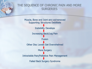
With typical laminotomy/discectomy, including the so-called minimally invasive techniques, bone and ligamentum flavum need to be removed in order to access the spinal canal and the pathology. Depending on the size and location of the extruded fragment/s, the amount of bone removal ranges from a conservative laminotomy to a wider laminectomy with or without hemi-facetectomy. In either case, once the normal anatomy has been altered the possibility of failed back surgical syndrome increases dramatically.
The use of a non-traumatic, small-sized, access tubular system allows for excellent visualization via scope viewing and working simultaneously in order to part through the ligamentum flavum. There is no internal cutting, bone or joint removal. Lack of bleeding virtually eliminates the risk of new scar tissue.
METHODS
A retrospective analysis from 2002 through the present was performed. The results are reported utilizing the MacNab criteria. The study population includes 111 patients with L5/S1 extruded disc herniations/free fragments into the spinal canal; 74% males and 26% females, ages ranging from 17 to 58 years old. The mean follow up was 6 weeks.
RESULTS
Statistical analysis showed the following results: Excellent: 57.65% (n=64), Good: 35.13% (n=39), Fair: 3.60% (n=4) and Poor: 3.60% (n=4). The overall success rate was 93%, or if the Fair result group is also included with modified MacNab criteria, it is 96%. One COMPLICATION was a small dural sac leak which occurred indirectly after removal of free fragments that were plastered against the dura. This was successfully treated with an epidural blood patch placed by the anesthesiologist; this patient was not hospitalized, had headaches with standing for two days that disappeared completely with the blood patch and the patient had an excellent outcome..
CONCLUSIONS
This method achieves better success rates and avoids the potentially deleterious long term ill effects of trauma that occur with typical procedures.

Presentation @ the Congress of Neurological Surgeons 2011 Annual Meeting, Oct 2011
INTRODUCTION
Lumbar fusion rates have steadily increased since the 1990’s. However, after lumbar fusion approximately 40% of patients remain unchanged or become even worse as measured by Oswestry at two year follow-up. Our analysis group represents those patients treated here who had fusion recommended, by outside surgeons, but the patients chose our smaller access out-patient discectomy instead. This procedure is a focused, precision discectomy. Our retrospective analysis goal was to determine whether these patients had undergone a later fusion. Our success is defined as those patients that have avoided fusion (90%).
METHODS
59 patients were available for follow-up. 35 % were women (n=21) and 65 % men (n=38) with an average age of 42 years old, ranging from 21 to 70 years of age. 16% of patients presented with primarily low back pain and 77% had objective signs of radiculitis. The average follow-up was 43 months.
RESULTS
There were 9 patients who underwent a reoperation: 6 fusions, 1 disc decompression, 1 laminectomy/foraminotomy and 1 minimally invasive discectomy. Therefore, our success rate for avoiding fusion was 90 % and for avoiding any further procedure was 85%. Of the 6 patients that required fusion: 1 had 2 previous lumbar operations, 1 had concomitant multiple sclerosis, 1 had a previous fusion one segment below and one had spondylolysthesis. All but 1 re-operated case presented with hard disks. 70 % of patients showed excellent and good clinical results. No complications were reported.
CONCLUSIONS
Fusion should only be done after a careful evaluation of the individual case taking into consideration all factors involved, including whether much smaller procedures may be sufficient. The use of the smaller access discectomy technique was shown above to be a valuable tool in avoiding fusion of the lumbar spine, even in cases where associated factors beyond the discopathy were not surgically treated.

Source: Presentation 2012 AANS/CNS Section on Disorders of the Spine and Peripheral Nerve Annual Meeting , Mar 2012. Orlando Florida.
INTRODUCTION
Multifactorial Spinal stenosis, whether central, lateral or foraminal, affects millions of Americans. The clinical presentation ranges from asymptomatic (radiological finding) to neurological claudication and radicular pain.
After exhausting conservative methods of treatment, most surgical solutions entail generous bone, ligament flavum, articular facet and disc removal. This in turn could generate, in a high percentage of cases, spinal instability which leads to fusion and possibly to failed back surgery syndrome.
The present abstract deals with the treatment of multifactorial spinal stenosis by treating one of the factors, the disc, with a focused lumbar discectomy.
METHODS
27 cases were obtained from a retrospective chart review from 2009 up to date. We included cases of patients older than 50 years of age with multifactorial spinal stenosis diagnosed by MRI and CT scan, interpreted by independent radiologists. All patients underwent CT scan after discography and were treated with a focused lumbar discectomy. Ages ranged from 50 to 86; 41% females and 59% males. 70 % of patients presented with a combination of central, recess and foraminal stenosis; 78% with radicular symptoms, 11% with lumbago and 11 % with neurological claudication, the latter in combination with radicular presentation. Average follow up was 8 wks.
RESULTS
Utilizing the MacNabb criteria the results were as follows: 41 % (n=11) excellent, 52% (n=14) good and 7% (n=2) Fair with a success rate of 93%. No complications were reported.
CONCLUSIONS
The treatment of multifactorial spinal stenosis with solely a primary factor approach lumbar discectomy is very effective and safe. At the same time it does not preclude the use of other more invasive surgical options should this approach be insufficient

The International Society for the Advancement of Spine Surgery in Boca Raton, FL USA | April 12 – April 14, 2017.
INTRODUCTION
Central disc herniations in the lumbar spine are difficult to access. Even with the so called “minimally invasive” techniques, substantial bone removal needs to occur which increases the likelihood of instability and failed back surgery syndrome in the long run.
We use a double, “nano” approach with an endoscope which produces no-access trauma to the normal spine in order to treat the pathology. The first access targets the center of the disc and provides decompression, the second access is more lateral and almost 90 degrees perpendicular to the A-P axis of the vertebral unit and targets specifically the herniated portion of the disc whether it is contained or extruded. The “offending” disc material is removed with the use of microtools. Because the second access is in close proximity to the abdominal cavity, it is essential to request a “full body thickness” CT through the affected levels in order to determine the safety of the access.
MATERIAL AND METHODS
A retrospective analysis from 2002 was performed by one of the authors. A total of 107 patients were gathered; 66% males and 34 % females, ages ranging 12 to 83 years old. There were 65 L4/5, 34 L5/S1 and 8 L3/4 herniations; 66% were contained and 34% extruded. The average f/u was 9 weeks. The results were reported utilizing the MacNab criteria.
RESULTS
No access limitation was showed on wide view CT scans. Results were as follows: Excellent: 52.33% (n=56), Good: 38.31% (n=41), Fair: 8.41% (n=9) and Poor: 0.95% (n=1) with an overall success rate of 90.64%. No complications were reported.
CONCLUSIONS
The term “minimally invasive” is very often used too liberally and with disregard for the actual changes the surgical procedure imposes on the normal spine. In that sense our “nano” procedure is the only minimally invasive spine procedure available today that causes no access trauma to the spine. This concept is specially true regarding central disc herniations because of their difficult access. Our technique is highly successful, very safe and avoids the deleterious long term effects of considerable bone removal and tissue dissection that may lead to failed back surgery syndrome.

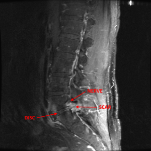
Some patients get MRI studies during the immediate period following a non-traumatic back procedure. Once performed, these MRI have the potential to confuse the entire clinical picture rather than to shed light about the healing process.
Many authors in the country and internationally have published important scientific papers about the characteristics of MRI changes in the postoperative of asymptomatic (no complaints) patients who had a successful lumbar disc operation. They found that there was no good correlation between symptoms and MRI images during that period. The MRI showed changes that mimic (look-like) the pre-surgical images; however, the patients were doing very well from the clinical standpoint. These MRI changes could sometimes persist for up to a year.
Scott Boden, MD published an article in Radiology, 1992, where he states that in the postoperative, the normal and typically expected enhancement pattern: “was indistinguishable from that seen with a recurrent (re-appearance) or a residual (the herniation was not taken out completely) disk fragment surrounded by scar or inflammatory tissue” and continues: “Decisions about surgery made on the basis of MR studies obtained during the first 3 to 6 months postoperatively must be carefully considered”.
Andrew Deutsch, MD (Spine 1993) performs MRI studies in 23 asymptomatic patients 1 year after successful lumbar disc operations. He found that 50 % of the patients showed posterior disc problems that mimicked the original herniated disc problem. He went on to describe 15% of cases in which no change or an even greater disc problem was shown in the MRI after a year of successful disc operation.
Even though these studies were performed in patients that did not have (or had minimal) symptoms, I wouldn’t like to give the false impression that post-surgical MRI studies are useless. Under special circumstances, they could add valuable information when ordered at the proper time. In conjunction with the clinical picture and an in-depth analysis of the actual films, these radiological studies could help the surgeon determine what is the best course of action for one’s particular case during the postoperative recovery.

In an era of tremendous technological advances in medicine, it is very tempting to “jump” to quick conclusions, diagnosis or recommendations solely based on high end technology like MRI. Many conservative and even surgical recommendations in patients with low back pain are based almost exclusively on abnormal MRI findings. However; some lumbar MRI findings do not correlate very accurately with the presence or not of low back pain.
In a study published by Jarvik in Spine in 2001 he reported MRI findings on 148 asymptomatic subjects. The results showed:
Clearly, many abnormal MRI findings were fairly common in patients without low back pain and were therefore of limited diagnostic value. However; findings of spinal stenosis, root compression and disc extrusions were diagnostically and clinically relevant despite being less common.
Nothing can replace the value of a thorough history and physical performed by an experienced specialist. The MRI evaluation coupled with the physical findings increases substantially the specificity and sensitivity of the MRI as a screening tool. In cases where the diagnosis is still in doubt, other studies such as discograms could be recommended to help shed light into the diagnostic dilemma.


