Practical Tips for Post-Spinal Surgery Recovery
Practical Tips for Post-Spinal Surgery Recovery

An orthopedic surgeon with more than 30 years of experience, Dr. Luis Lombardi dedicates his professional life to the development and improvement of spine surgical techniques that  do not damage the normal structures. In a recent interview,  Dr. Luis Lombardi stated “these non-traumatic procedures are performed in an outpatient setting, offer a shorter recovery times and require no anesthesia”. Even though these are small procedures that are extremely well tolerated by patients, you may want to take the following tips into account after any spine surgery. 1. Arrange for someone to transport you to and from the ambulatory center. 2. Make sure to attend all follow-up appointments with your orthopedic physician after the procedure. He can help monitor your recovery, address concerns and offer suggestions for recovery. 3. The success of any surgical procedure partly depends on the patient’s cooperation with pre and post-surgery instructions. Post-procedure management is important, so make sure to closely follow all instructions given to you by the surgeon. 4. Your orthopedic surgeon may advise you to avoid excessive bending and heavy lifting within the first few days after your procedure.

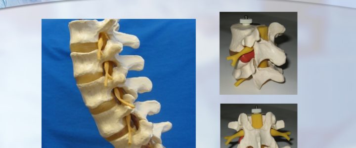
Anatomy of the Vertebra and Disc by Dr. Luis Lombardi

Anatomy of the Vertebra and Disc by Dr. Luis Lombardi

Since 2000, Dr. Luis Lombardi has served as a privately practicing non-traumatic spine surgeon in Los Angeles, California. Dr. Luis Lombardi draws on a comprehensive knowledge of both normal and abnormal vertebral anatomy. The human spine is made up of 33 interlocking vertebrae, 24 of which are independently mobile. Each of these vertebrae consists of a cylindrical solid structure known as the vertebral body, which can withstand body weight and bears compression generated from pressure and movement. On the interior side of this structure lies the vertebral arch, which together with the vertebral body makes a circle around the spinal canal. The vertebral arch connects to a series of bony processes, to which the skeletal muscles connect. In between each pair of vertebrae lies the intervertebral disc. The disc consists of a shock-absorbing nucleus, which is contained within a harder protective annulus. The exterior third of the annulus contains pain-sensitive nerve fibers that activate in instances of disc damage. The primary nerves in the spine, however, pass down the spine behind the vertebrae and discs. Located close to the interior edge of the disc, the nerves branch out to become nerve roots, which come together to become the larger nerves of the legs

Causes and Symptoms of Sciatica by Dr Luis Lombardi

Causes and Symptoms of Sciatica by Dr Luis Lombardi

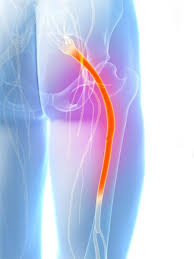
As a non-traumatic spine surgeon, Dr. Luis Lombardi employs a minimally invasive approach that targets the cause of a patient’s pain without unnecessary damage to surrounding structures. Dr. Luis Lombardi applies this treatment methodology to a range of spinal conditions, including sciatica. The term sciatica refers not to a diagnosable medical condition, but rather to a set of symptoms that indicate an underlying issue. Such symptoms typically stem from an irritation or compression of the sciatic nerve in the lower back. This often occurs when an intervertebral disc in the lower spine becomes worn and herniates. This causes the soft core of the disc to leak out through its protective coating and encroach on the surrounding nerves, including the sciatic nerve. The sciatic nerve, the longest in the human body, begins in the lower back and then splits into two. Each branch runs through a buttock, hip, leg, and feet. For this reason, when the sciatic nerve becomes compressed, patients experience pain that begins in the back and radiates down part or all of the long nerve. Patients typically describe the pain not as an ache but as a sharp, burning, or tingling discomfort. It may co-present with weakness or numbness in the same leg and may cause difficulty walking, though the pain itself is often worse when sitting.

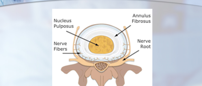
Anatomy of the Vertebra and Intervertebral Disc by Dr. Luis Lombardi

Anatomy of the Vertebra and Intervertebral Disc by Dr. Luis Lombardi

Since 2000, Dr. Luis Lombardi has served as a privately practicing non-traumatic spine surgeon in Los Angeles, California. Dr. Luis Lombardi draws on a comprehensive knowledge of both normal and abnormal vertebral anatomy.

The human spine is made up of 33 interlocking vertebrae, 24 of which are independently mobile. Each of these vertebrae consists of a cylindrical solid structure known as the vertebral body, which can withstand body weight and bears compression generated from pressure and movement. On the interior side of this structure lies the vertebral arch, which together with the vertebral body makes a circle around the spinal canal. The vertebral arch connects to a series of bony processes, to which the skeletal muscles connect. In between each pair of vertebrae lies the intervertebral disc. The disc consists of a shock-absorbing nucleus, which is contained within a harder protective annulus. The exterior third of the annulus contains pain-sensitive nerve fibers that activate in instances of disc damage. The primary nerves in the spine, however, pass down the spine behind the vertebrae and discs. Located close to the interior edge of the disc, the nerves branch out to become nerve roots, which come together to become the larger nerves of the legs.


Some Basic Spinal Care Tips by Dr. Luis Lombardi

Some Basic Spinal Care Tips by Dr. Luis Lombardi

Here are some basic spinal care tips:

– Sleeping posture is vital in spinal care. As much as possible, experts recommend sleeping on the back, though sleeping on the sides is also acceptable. However, sleeping on the stomach should be avoided as it imparts excess pressure on the spine. Using a supportive mattress and a pillow that aligns the neck properly are also important to providing complete rest to the spine while sleeping.

– Wearing the right pair of shoes can help support the back as well. Shoes that fit perfectly, that check pronation or supination (or roll) of the foot and provide a platform that helps to align the spine and the body are recommended.

– Spinal care also means paying attention to problems like back pain, hip pain, or weaknesses in the spine. Experiencing these issues once in a while is common, but they also could be symptoms of serious spinal problems. It is not a good idea to self-medicate to relieve these symptoms or over stress the body by doing physical activities like a workout. It is better to consult a medical professional to treat the symptoms over time.

Dr Luis Lombardi-Postoperative MRI

Dr Luis Lombardi-Postoperative MRI

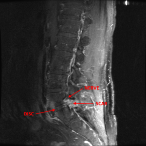
Some patients get MRI studies during the immediate period following a non-traumatic back procedure. Once performed, these MRI have the potential to confuse the entire clinical picture rather than to shed light about the healing process.

Many authors in the country and internationally have published important scientific papers about the characteristics of MRI changes in the postoperative of asymptomatic (no complaints) patients who had a successful lumbar disc operation. They found that there was no good correlation between symptoms and MRI images during that period. The MRI showed changes that mimic (look-like) the pre-surgical images; however, the patients were doing very well from the clinical standpoint. These MRI changes could sometimes persist for up to a year.

Scott Boden, MD published an article in Radiology, 1992, where he states that in the postoperative, the normal and typically expected enhancement pattern: “was indistinguishable from that seen with a recurrent (re-appearance) or a residual (the herniation was not taken out completely) disk fragment surrounded by scar or inflammatory tissue” and continues:  “Decisions about surgery made on the basis of MR studies obtained during the first 3 to 6 months postoperatively must be carefully considered”.

Andrew Deutsch, MD (Spine 1993) performs MRI studies in 23 asymptomatic patients 1 year after successful lumbar disc operations. He found that 50 % of the patients showed posterior disc problems that mimicked the original herniated disc problem. He went on to describe 15% of cases in which no change or an even greater disc problem was shown in the MRI after a year of successful disc operation.

Even though these studies were performed in patients that did not have (or had minimal) symptoms, I wouldn’t like to give the false impression that post-surgical MRI studies are useless. Under special circumstances, they could add valuable information when ordered at the proper time. In conjunction with the clinical picture and an in-depth analysis of the actual films,  these radiological studies could help the surgeon determine what is the best course of action for one’s particular case during the postoperative recovery.

Dr. Luis Lombardi-MRI FINDINGS IN LOW BACK PAIN

Dr. Luis Lombardi-MRI FINDINGS IN LOW BACK PAIN

In an era of tremendous technological advances in medicine, it is very tempting to “jump” to quick conclusions, diagnosis or recommendations solely based on high end technology like MRI. Many conservative and even surgical recommendations in patients with low back pain are based almost exclusively on abnormal MRI findings. However; some lumbar MRI findings do not correlate very accurately with the presence or not of low back pain.

In a study published by Jarvik in Spine in 2001 he reported MRI findings on 148 asymptomatic subjects. The results showed:

  • 83% with moderate to severe desiccation of one or more discs
  • 64% with one or more bulging discs.
  • 56% with loss of disc height.
  • 32% had at least one disc protrusion
  • 6% had one or more disc extrusions

Clearly, many abnormal MRI findings were fairly common in patients without low back pain and were therefore of limited diagnostic value. However; findings of spinal stenosis, root compression and disc extrusions were diagnostically and clinically relevant despite being less common.

Nothing can replace the value of a thorough history and physical performed by an experienced specialist. The MRI evaluation coupled with the physical findings increases substantially the specificity and sensitivity of the MRI as a screening tool. In cases where the diagnosis is still in doubt, other studies such as discograms could be recommended to help shed light into the diagnostic dilemma.

Dr Luis Lombardi-Testimonial

Dr Luis Lombardi-Testimonial
“ Last January, I took a bad fall on the ice resulting in constant sciatica. An MRI revealed two herniated discs. I had two spinal injections, physical therapy, chiropractic care-none of which helped. I couldn’t sit, stand or sleep without extreme pain. Two different surgeons from well-known hospitals told me I had no choice but traditional surgery which will also include fusion…. I traveled from Illinois to California and had my procedure with Dr. Luis Lombardi….the results were nothing short of miraculous. My husband was skeptic before the surgery and now he is a believer. He had to practically carry me into the clinic before the procedure because I could barely walk. When I awoke afterwards, I had no pain. It was the best feeling in the world. That evening, we went out to dinner and afterwards I had my first night of sleep with no pain in 8 months. It is now over 2 years later and I am walking several miles every morning and able to care for my family again. Every time I think about all of this, I consider myself the most fortunate person to have found you. “
Lynne A.

Glen Ellyn, IL

Dr. Luis Lombardi-Testimonial Lynne A.

“ Last January, I took a bad fall on the ice resulting in constant sciatica. An MRI revealed two herniated discs. I had two spinal injections, physical therapy, chiropractic care-none of which helped. I couldn’t sit, stand or sleep without extreme pain. Two different surgeons from well-known hospitals told me I had no choice but traditional surgery which will also include fusion…. I traveled from Illinois to California and had my procedure with Dr. Luis Lombardi….the results were nothing short of miraculous. My husband was skeptic before the surgery and now he is a believer. He had to practically carry me into the clinic before the procedure because I could barely walk. When I awoke afterwards, I had no pain. It was the best feeling in the world. That evening, we went out to dinner and afterwards I had my first night of sleep with no pain in 8 months. It is now over 2 years later and I am walking several miles every morning and able to care for my family again. Every time I think about all of this, I consider myself the most fortunate person to have found you. “

Lynne A.